A study published in Radiology: Cardiothoracic Imaging evaluated the chest CT findings of 104 cases with COVID-19 from the cruise ship 'Diamond Princess'.
Abstract
Purpose
To evaluate the chest CT findings in an environmentally homogeneous cohort from the cruise ship “Diamond Princess” with Coronavirus Disease 2019 (COVID-19).
Materials and Methods
This retrospective study comprised 104 cases (mean age, 62 years ± 16, range 25-93) with COVID-19 confirmed with RT-PCR. CT images were reviewed and the CT severity score was calculated for each lobes and the entire lung. CT findings were compared between asymptomatic and symptomatic cases.
Results
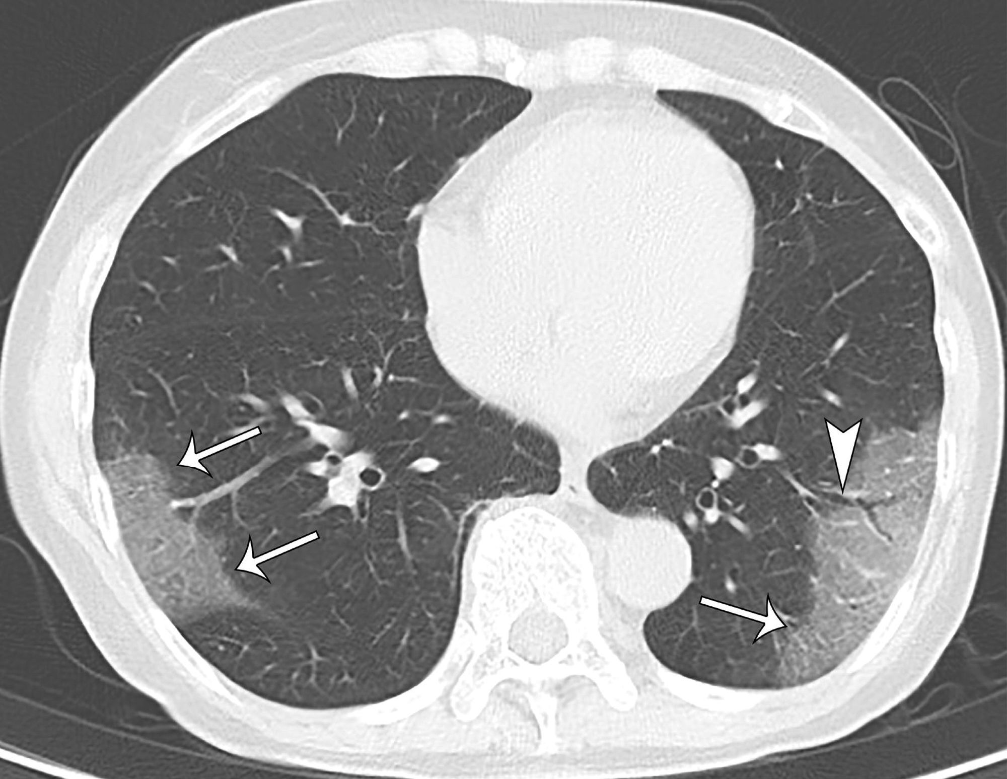
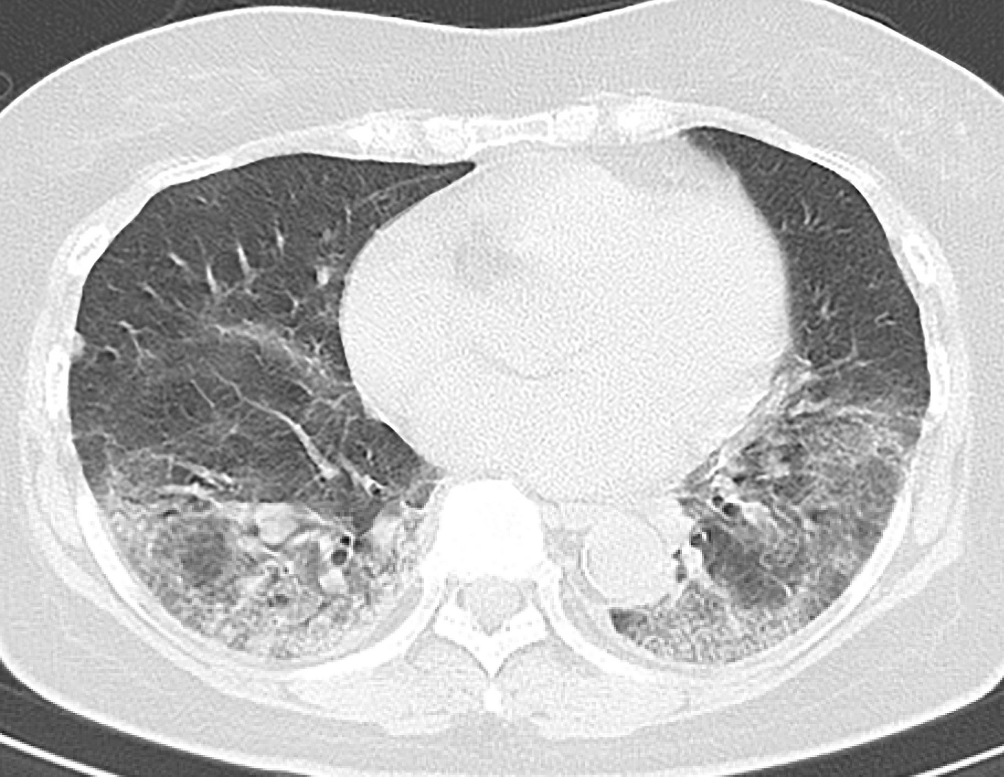
Of 104 cases, 76 (73%) were asymptomatic, 41 (54%) of which had lung opacities on CT. Other 28 (27%) cases were symptomatic, 22 (79%) of which had abnormal CT findings. Symptomatic cases showed lung opacities and airway abnormalities on CT more frequently than asymptomatic cases [lung opacity; 22 (79%) vs 41 (54%), airway abnormalities; 14 (50%) vs 15 (20%)]. Asymptomatic cases showed more GGO over consolidation (83%), while symptomatic cases more frequently showed consolidation over GGO (41%). The CT severity score was higher in symptomatic cases than asymptomatic cases, particularly in the lower lobes [symptomatic vs asymptomatic cases; right lower lobe: 2 ± 1 (0-4) vs 1 ± 1 (0-4); left lower lobe: 2 ± 1 (0-4) vs 1 ± 1 (0-3); total score: 7 ± 5 (1-17) vs 4 ± 2 (1-11)].
Conclusion
This study documented a high incidence of subclinical CT changes in cases with COVID-19. Compared to symptomatic cases, asymptomatic cases showed more GGO over consolidation and milder extension of disease on CT.
Summary
We revealed a high incidence of subclinical CT changes in COVID-19 infected cases, which showed more GGO predominance over consolidation and milder severity on CT than symptomatic cases.
Key Points
■ Of 104 cases analyzed, 76 (73%) were asymptomatic, 41 (54%) of which had pneumonic changes on CT. Other 28 (27%) cases were symptomatic, 22 (79%) of which had abnormal CT findings.
■ Asymptomatic cases showed more GGO predominance over consolidation (83%), while symptomatic cases were more likely to show a consolidation predominance over GGO (41%).
■ Asymptomatic cases showed milder CT severity score than symptomatic cases.
Chest CT Findings in Cases from the Cruise Ship “Diamond Princess” with Coronavirus Disease 2019 (COVID-19)Shohei Inui, Akira Fujikawa, Motoyuki Jitsu, Naoaki Kunishima, Sadahiro Watanabe, Yuhi Suzuki, Satoshi Umeda, and Yasuhide UwabeRadiology: Cardiothoracic Imaging20202:2 https://doi.org/10.1148/ryct.2020200110